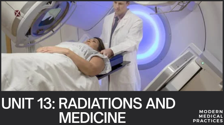
PHYSICS S6 UNIT 13: RADIATIONS AND MEDICINE.

About Course
Radiation plays a vital and increasingly sophisticated role in modern medicine, encompassing both diagnostic and therapeutic applications. It’s a field that has revolutionized our ability to see inside the human body, detect diseases early, and treat various conditions, particularly cancer.
Here’s a breakdown of the key aspects of radiations and medicine:
I. Types of Radiation Used in Medicine:
The primary types of radiation used are:
-
Ionizing Radiation: This is the most common type used in medical imaging and treatment. It has enough energy to remove electrons from atoms, creating ions. This process can damage DNA and cells, which is why its use needs to be carefully controlled.
-
X-rays: A form of electromagnetic radiation used for diagnostic imaging (radiography, CT scans, mammography). They pass through soft tissues but are absorbed by denser materials like bone, creating a shadow image.
-
Gamma Rays: Also a form of electromagnetic radiation, often emitted from radioactive isotopes (radioisotopes). Used in nuclear medicine for both diagnostic imaging (SPECT, PET scans) and therapy.
-
Alpha Particles: Consist of two protons and two neutrons (like a helium nucleus). They are heavy and highly ionizing but have a very short range. Primarily used in targeted alpha therapy (TAT) for very localized cancer treatment.
-
Beta Particles: High-energy electrons or positrons. They have a longer range than alpha particles but are still relatively short. Used in some nuclear medicine therapies (e.g., radioactive iodine for thyroid conditions).
-
Protons and other charged particles: Used in advanced radiation therapy techniques (e.g., proton therapy) due to their precise dose delivery characteristics, which can spare surrounding healthy tissue.
-
-
Non-Ionizing Radiation: While not typically categorized as “radiation” in the context of X-rays or nuclear medicine, other forms of electromagnetic radiation are also used:
-
Radiofrequency (RF) waves: Used in Magnetic Resonance Imaging (MRI) to create detailed images of soft tissues.
-
Ultraviolet (UV) light: Used for sterilization of medical equipment and in some dermatological treatments.
-
Laser light: Used in various surgical procedures (e.g., ophthalmology, dermatology) and for diagnostic purposes.
-
II. Diagnostic Applications (Medical Imaging):
The goal here is to visualize internal structures or assess organ function without invasive surgery.
-
Radiography (X-rays):
-
Standard X-rays: Used to detect bone fractures, lung conditions (pneumonia, emphysema), dental issues, etc.
-
Mammography: Specialized X-ray for breast cancer screening.
-
Fluoroscopy: Real-time X-ray imaging, often with a contrast agent, to visualize dynamic processes (e.g., blood flow, digestive tract movement).
-
-
Computed Tomography (CT Scan):
-
Combines multiple X-ray images from different angles to create cross-sectional (slice) images of the body. Provides much more detailed anatomical information than standard X-rays. Used for diagnosing a wide range of conditions, including tumors, internal injuries, and vascular problems.
-
-
Nuclear Medicine (using Radioisotopes/Radiopharmaceuticals):
-
Patients are administered a small amount of a radioactive tracer (radiopharmaceutical) that targets specific organs or tissues. The tracer emits gamma rays, which are detected by special cameras.
-
SPECT (Single-Photon Emission Computed Tomography): Creates 3D images that show blood flow and organ function.
-
PET (Positron Emission Tomography): Uses positron-emitting radioisotopes to create detailed functional images, particularly useful for detecting cancer, assessing brain activity, and evaluating heart disease.
-
-
Magnetic Resonance Imaging (MRI):
-
While not using ionizing radiation, MRI is a powerful imaging technique that uses strong magnetic fields and radio waves to produce highly detailed images of soft tissues, organs, bone, and virtually all other internal body structures. Excellent for brain, spinal cord, joint, and abdominal imaging.
-
III. Therapeutic Applications (Radiation Therapy/Radiotherapy):
The primary goal is to destroy diseased cells, especially cancer cells, while minimizing damage to healthy tissue.
-
External Beam Radiation Therapy (EBRT):
-
Radiation is delivered from a machine outside the body. This is the most common type of radiation therapy.
-
3D Conformal Radiation Therapy (3D-CRT): Shapes the radiation beams to conform to the shape of the tumor.
-
Intensity-Modulated Radiation Therapy (IMRT): Allows for even more precise shaping and intensity variation of the radiation beams, delivering higher doses to the tumor while sparing critical organs.
-
Image-Guided Radiation Therapy (IGRT): Uses imaging (e.g., daily CT scans) to precisely locate the tumor before each treatment, accounting for patient and tumor movement.
-
Stereotactic Radiosurgery (SRS) / Stereotactic Body Radiation Therapy (SBRT): Delivers a very high dose of radiation in a few fractions (or even a single fraction) with extreme precision to small tumors.
-
Proton Therapy: Uses protons instead of X-rays. Protons deposit most of their energy at a specific depth (Bragg peak), allowing for precise dose delivery with minimal exit dose, beneficial for tumors near sensitive structures.
-
-
Brachytherapy (Internal Radiation Therapy):
-
Radioactive sources are placed directly inside or very close to the tumor.
-
Temporary Brachytherapy: Sources are placed for a specific period and then removed (e.g., for prostate or cervical cancer).
-
Permanent Brachytherapy: Small radioactive “seeds” are implanted permanently and deliver radiation over time (e.g., for prostate cancer).
-
-
Systemic Radiation Therapy (Nuclear Medicine Therapy):
-
Radioactive substances are given orally or intravenously and travel throughout the body to target specific cells or tissues.
-
Radioactive Iodine (I-131): Used to treat thyroid cancer and hyperthyroidism because thyroid cells absorb iodine.
-
Radium-223 (Xofigo): Used for metastatic prostate cancer that has spread to bones.
-
Lutetium-177 (Lu-177 DOTATATE): Used for neuroendocrine tumors.
-
Course Content
RADIATION DOSE.
-
Ionization and non-ionization radiations.
23:41 -
Radiation penetration in body tissue.
21:20 -
Radiation dosimetry.
25:12 -
Radiation exposure.
19:23 -
Absorbed radiation dose.
18:16 -
Quality Factors.
14:46 -
Equivalent Dose.
12:15 -
Radiation Protection.
20:42 -
Checking my Progress.
29:17 -
Questions and Answers.
44:21
BIOLOGICAL EFFECTS OF RADIATION EXPOSURE.
CONCEPT OF BALANCED RISK.
THE HALF-LIVES: PHYSICAL, BIOLOGICAL, AND FFECTIVE.
END UNIT ASSESSMENT
Questions and Answers.
Final Unit Exam.
Student Ratings & Reviews
